Overview
The Microscopic Imaging Platform delivers comprehensive optical microscopy access services to meet diverse research demands of the Institute of Immunology, as well as institutions inside and outside the university in microscopic imaging, image analysis and processing, and related fields. The platform is equipped with a full array of imaging instruments including two-photon microscopes, laser scanning confocal microscopes, whole-slide digital scanning systems, light-sheet microscopes, stereoscopic fluorescence microscopes, upright and inverted fluorescence microscopes, alongside dedicated workstations installed with professional image analysis software. Moreover, it provides sample preparation services using a vibrating microtome, cryostat and microwave rapid tissue processor, as well as surgical procedures prior to in vivo imaging of animal tissues.
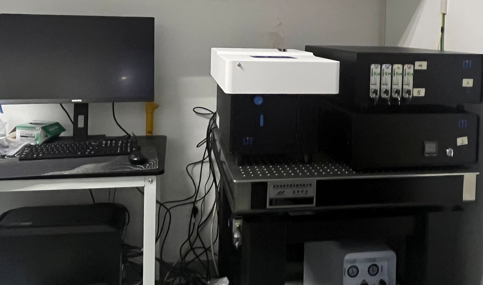
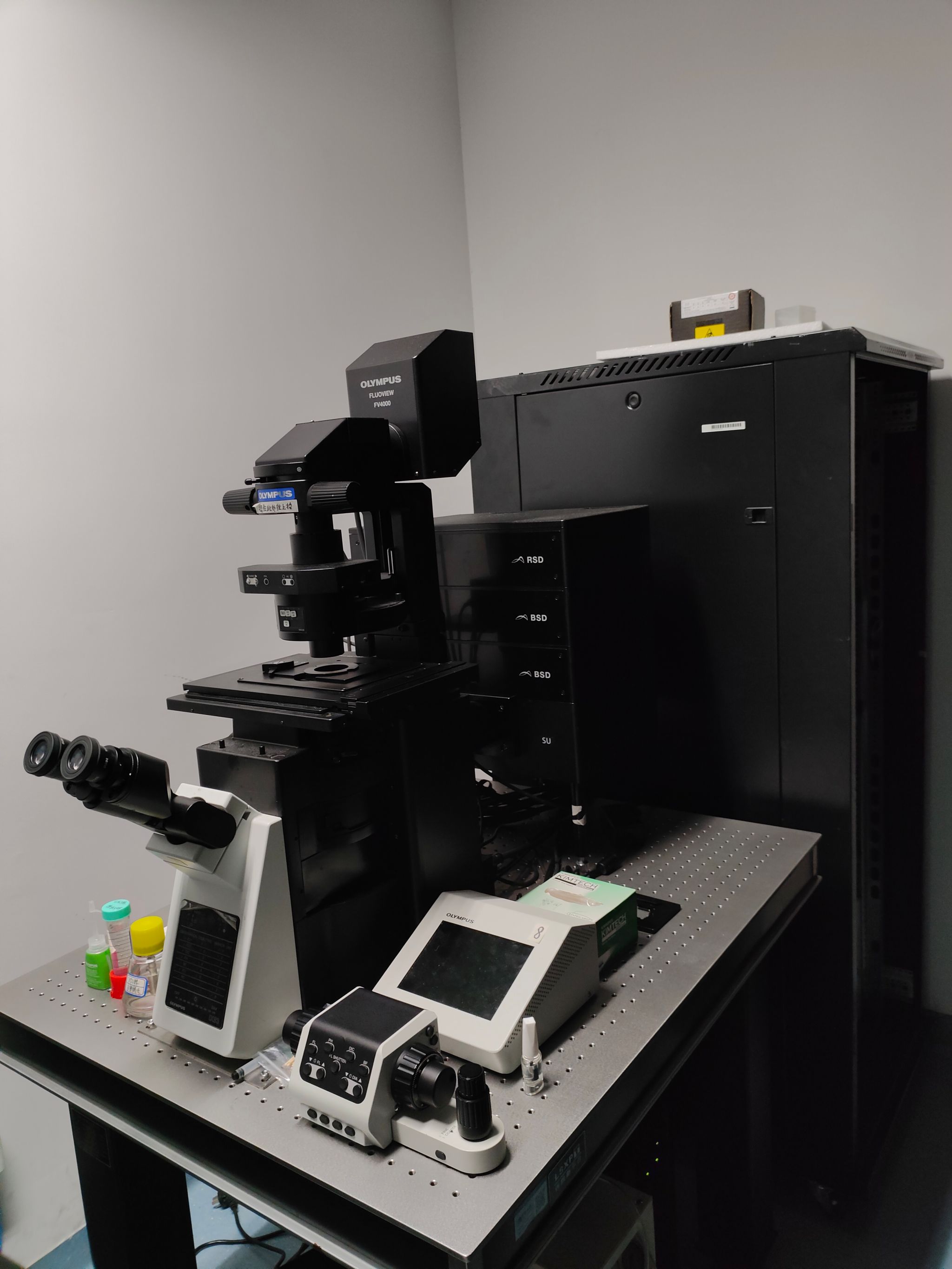
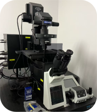
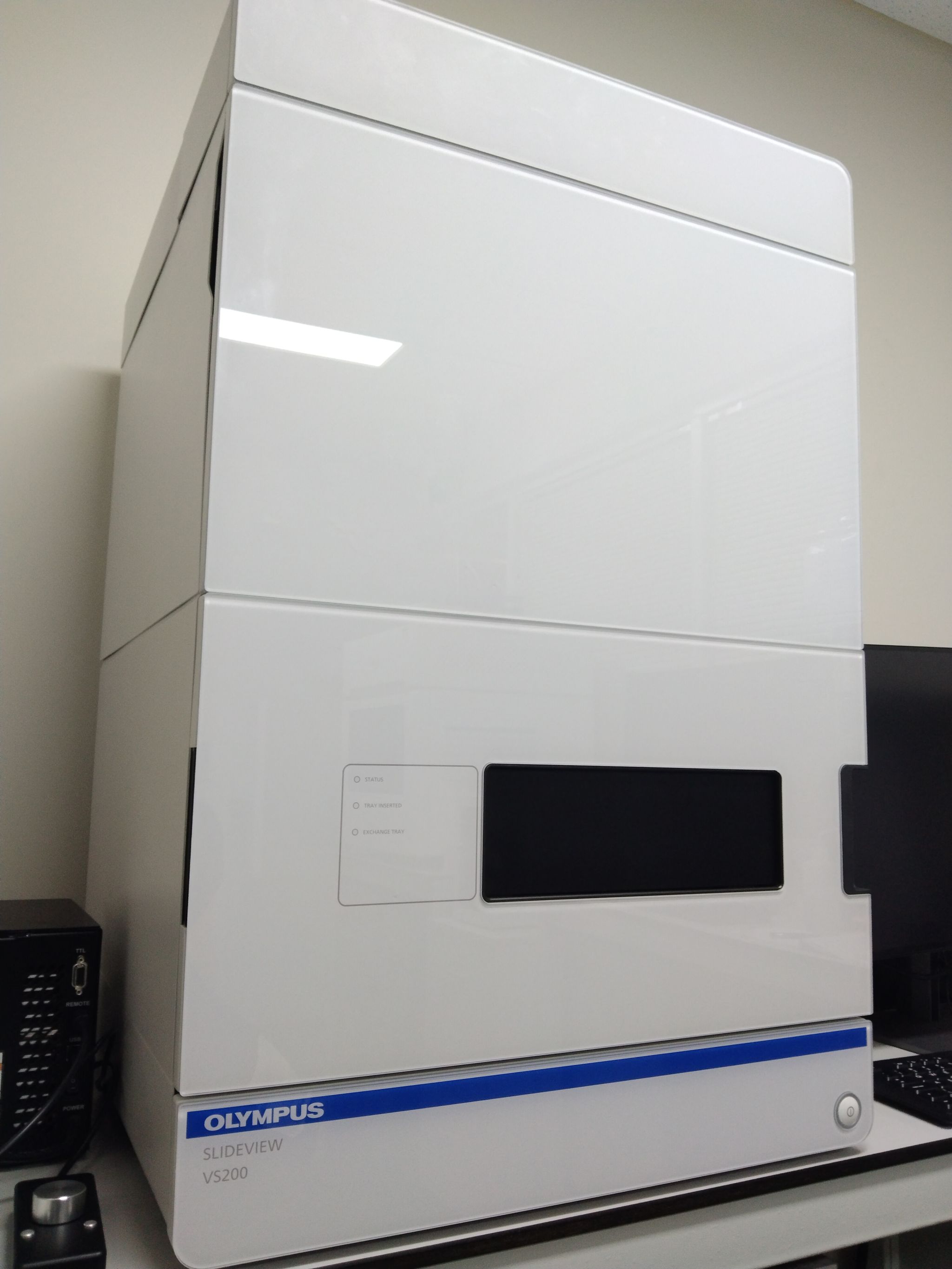
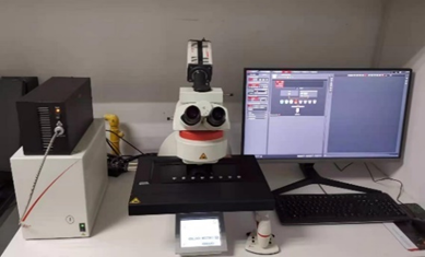
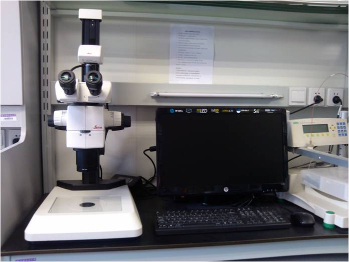
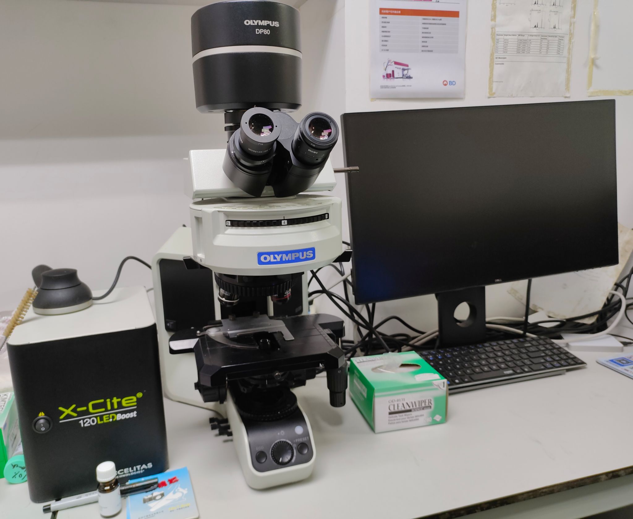
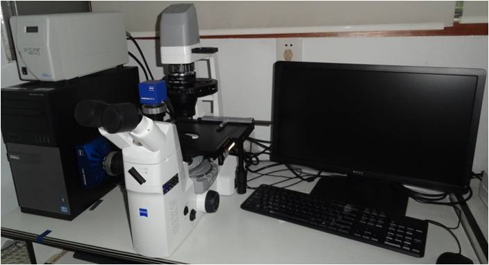
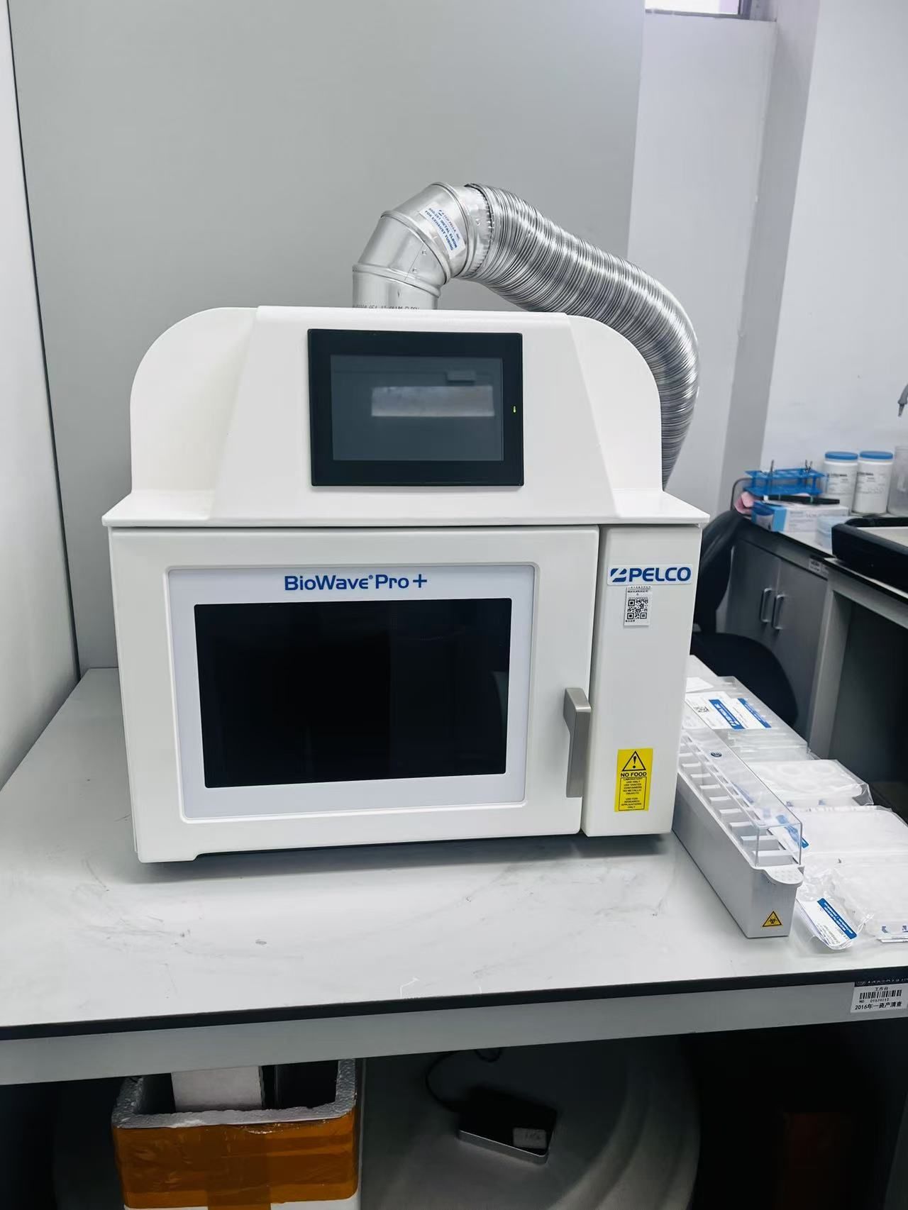
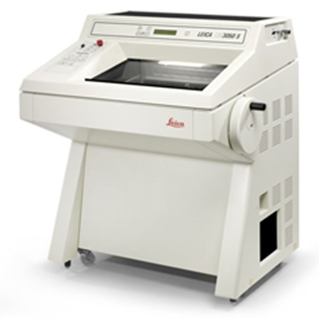
The platform currently houses the following instruments:1 unit of two-photon microscope (Olympus FVMPE-RS);4 laser scanning confocal microscopes (SpinSR10 spinning-disk super-resolution microscope, Olympus FV4000, Olympus FV3000, Leica SP8);2 whole-slide scanning systems (VS200, Leica THUNDER DM6);1 light-sheet microscope (LiTone XL);1 high-resolution fully automated stereoscopic fluorescence microscope (Leica THUNDER Imager M205FA);1 standard upright microscope, 1 standard inverted microscope and 1 standard stereomicroscope, respectively.In addition, the platform is furnished with 1 vibrating microtome (Leica VT1200S), 1 cryostat (Leica CM3050S), 1 microwave rapid tissue processor (PELCO BioWave Pro+), and 2 data analysis workstations pre-installed with professional software such as Imaris and HALO.
Equipment
OLYMPUS FLUOVIEW FVMPE-RS |
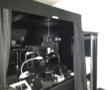
LiTone XL |
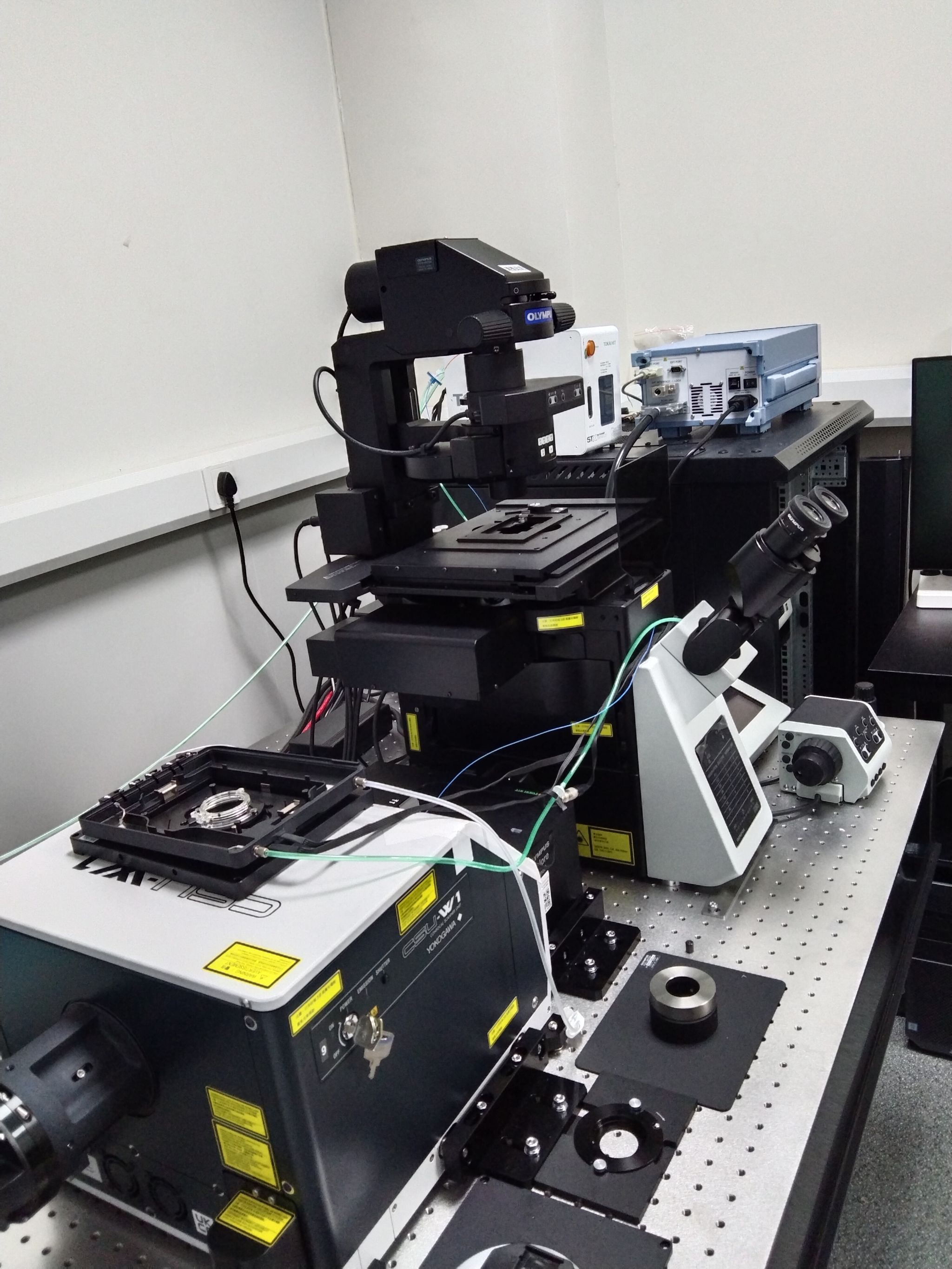
OLYMPUS IXPLORE SPINSR10 |
OLYMPUS FLUOVIEW FV3000(Upgraded Version ) |
OLYMPUS FLUOVIEW FV3000(1209 ) |
Leica SP8 |
OLYMPUS SLIDEVIEW VS200 |
THUNDER DM6 |
|
|
Zeiss Axio Vert A1 |
Image Analysis Workstation |
PELCO BioWave Pro+ |
Leica VT1200S |
LeicaCM3050S |
Service
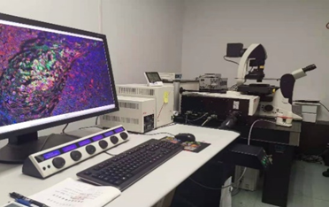
The platform fulfills multi-dimensional microscopic imaging requirements covering the full workflow from tissue sectioning, image acquisition to data analysis. It offers a complete range of technical services, including multicolor fluorescence two-dimensional (X,Y) and three-dimensional (X,Y,Z) imaging, time-lapse scanning, fully automated large-area image stitching, whole-slide scanning, large specimen imaging, cellular super-resolution imaging, live-cell dynamic image capture, and in vivo observation of model organisms.The platform supports visualization, analysis, segmentation and interpretation of various 2D/3D/4D microscopic images. It enables quantitative measurement on target objects, dynamic tracking of multiple targets, multi-dimensional fluorescence localization and other analytical operations. Furthermore, it can process panoramic scanned images of multiplex immunohistochemistry-stained sections, conducting tissue region partitioning, cell identification, fluorescence quantification, spatial relationship assessment and other downstream analyses.
In vivo microscopic imaging services for animal tissues are available on the platform, with a set of mature, validated protocols established for in vivo imaging of nearly all tissues and organs in mice. The platform delivers an annual average of over 10,000 instrument service hours, supporting more than 20 research groups within the institute, as well as over 10 external research groups and affiliated institutions.
Contact us
Director:
Dr.Jing Wang (jingwangATshsmu.edu.cn)
Staff:
Wei Jin (woshijinweiaAT126.com)
Wenjuan Bai (wjbaiATshsmu.edu.cn)
Yongxiao Liu (liuyongxiaoATshsmu.edu.cn)