On August 25th, 2023, A Frontal Transcallosal Inhibition Loop Mediates Interhemispheric Balance in Visual is published online in the journal Nature Communications The study was jointly completed by Zhang Siyu's research team from the School of Medicine of Shanghai Jiao Tong University and Xu Min's research team from the Center of Excellence in Brain Science and Intelligent Technology (Institute of Neuroscience) of the Chinese Academy of Sciences. The study was based on the detection behavior of two-choice unforced visual changes in mice, using a combination of single-cell calcium imaging, photogenetics and brain slice electrophysiology. It was found that there is a inhibitory pathway between ACA in the left and right anterior cingulate cortex, which is composed of pyramid-neuron CPN that projects across the corpus callosum and PV+cal neuron that receives the project across the corpus callosum. This loop controls the bilateral hemispheric inhibition balance between the corpus callosum and thus plays an important role in visuospatial information processing. In the case of damage to one prefrontal lobe, enhancing the activity of PV+ neurons in the damaged contralateral prefrontal lobe to rebalance bilateral interhemispheric activity can correct visuospatial preference caused by damage, similar to the "Sprague effect" observed in primates, suggesting that the anterior cingulate gyrus is mainly involved in components related to visual selective attention in the detection of visuospatial changes.
The exchange of information between the two brain hemispheres is essential for processing sensory information, and the corpus callosum, as the largest white matter structure in the brain, connects many brain regions [1]. Previous studies have found that the transcallosal inhibition imbalance caused by frontal parietal cortex injury will break the activity balance between the two sides of the brain, and in severe cases, animals will ignore the visual stimuli in the space opposite to the injury [2]. However, the neural circuitry underlying this phenomenon remains unclear.
In order to explore this problem, researchers improved the task of detecting changes in visual stimuli [3] by placing visual stimuli in the bilateral monocular region of mice, which is conducive to detecting the interaction between bilateral hemispheres. Then, single-cell calcium imaging technology was used to record the CPN activity of trans-callosal projective cone neurons in the cingulate cortex region of mice during the task (Figure 1).
The recording results showed that the number of CPNS that preferred to respond to contralateral visual stimuli was significantly higher than that of those that preferred to respond to ipsilateral stimuli, and the responses of all CPNS recorded on average were stronger in contralateral tasks than in ipsilateral tasks, indicating that CPNS were mainly responsible for processing visual information in contralateral space. Photogenetic activation of CPN caused contralateral behavior to improve, ipsilateral behavior to deteriorate, and inhibition to obtain the opposite result (Figure 2), suggesting that CPN regulates the bilateral hemispheric ACA competitive interaction.
Inhibition of CPN in the axon endings of the contralateral ACA is similar to the behavioral changes induced by inhibition of these CPN cells, suggesting that transcallosal projection is the main pathway for CPN in ACA to regulate this behavior (Figure 3).
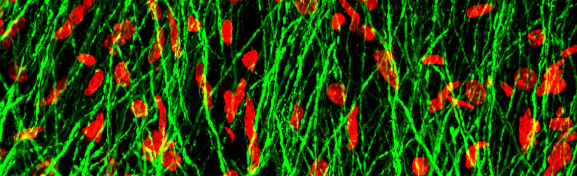
In order to further investigate which types of interneurons inhibit contralateral excitatory neurons by CPN, pseudorabies virus reverse tracing technique is used and it is found that excitatory neurons in contralateral ACA brain region and three main interneurons (PV+, SST+ and VIP+ neurons) are innervated by CPN. Electrophysiological results of brain film showed that CPNs selectively suppressed CPN in the contralateral ACA brain region mainly through PV+ neurons, especially PV+cal neurons that received strong input across the corpus callosum, thus forming a inhibitory loop across the corpus callosum (Figure 4).
More importantly, a temporary increase in the activity of PV+ neurons in the damaged antiACA region of unilateral ACA damaged mice (only during the task learning phase) can reverse the visuospatial preference caused by unilateral ACA damage and improve the visual change detection performance of the damaged antiACA in the long term (Figure 5). In clinical practice, some stroke patients with unilateral frontal parietal visual selective attention network impairment show hemispherical spatial neglect, which ignores the visual stimulation of the damaged opposite side. This work suggests that enhancing trans-callosal inhibition of the damaged opposite side may be a new idea for the treatment of corresponding attention deficit diseases.
The study was conducted by doctoral students Yanjie Wang and Zhaonan Chen and associate researcher Guofen Ma under the guidance of Siyu Zhang, School of Medicine, Shanghai Jiao Tong University, and Min Xu, Center of Excellence for Brain and Intelligence, Chinese Academy of Sciences. Doctoral student Wang Lizhao, postdoctoral student Liu Yanmei, undergraduate student Wu Yifan, senior experimentalist Qin Meiling and doctoral student Fei Xiang of the Center of Excellence also made important contributions. The work was funded by the Science and Technology Innovation 2030 Project of the Ministry of Science and Technology, the National Natural Science Foundation of China, the City of Shanghai, and the Chinese Academy of Sciences.
Reference
[1] INNOCENTI G, SCHMIDT K, MILLERET C, et al. The functional characterization of callosal connections [J]. Progress in neurobiology, 2022, 208: 102186.
[2] CORBETTA M, SHULMAN G. Spatial neglect and attention networks [J]. Annual review of neuroscience, 2011, 34: 569-99.
[3] BURGESS C P, LAK A, STEINMETZ N A, et al. High-Yield Methods for Accurate Two-Alternative Visual Psychophysics in Head-Fixed Mice [J]. Cell Rep, 2017, 20(10): 2513-24.


 Location:
>
Location:
>